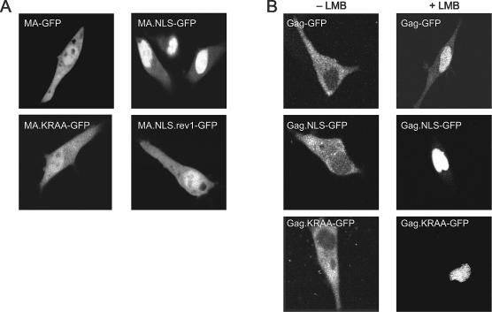
FIG. 2.
Subcellular localization of wild-type and mutant MA-GFP and Gag-GFP fusion proteins. (A) Live QT6 cells expressing wild-type or mutant MA-GFP fusion proteins were examined by fluorescence confocal microscopy 18 to 24 h after transfection. (B) QT6 cells transfected with plasmid DNAs expressing wild-type or mutant Gag-GFP fusion proteins were either untreated (−LMB) or treated (+LMB) with 20 nM LMB for 2 h and examined by confocal microscopy.

