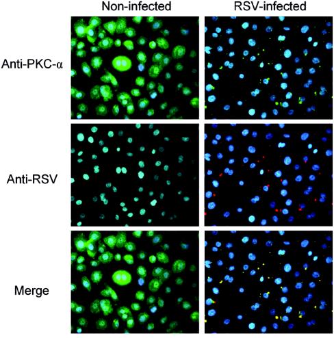
FIG. 6.
PKC-α colocalizes with RSV at early stages of infection. Confluent NHBE cells grown on 8-well chamber slides were incubated with 20 MOI of RSV for 1 h at 4°C to allow viral binding synchronization. Slides were then exposed at 37°C for 10 min to allow virus penetration. Later, cells were processed for immunocytofluorescence. Cells were fixed with 4% paraformaldehyde, permeabilized with 0.1% saponin, and stained with mouse monoclonal anti-PKC-α antibody (green), goat polyclonal anti-RSV antibody (red), and DAPI (blue, nucleus staining). Fluorescence images (magnification, ×400) were taken by cooled camera device under respective dual-filter mode (either green-blue or red-blue) and triple-filter mode (merge).

