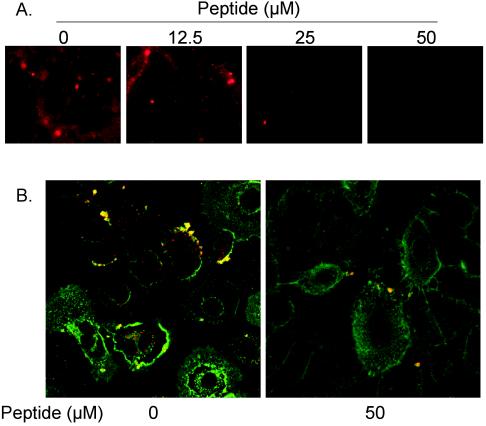
FIG. 8.
(A) Inhibition of PKC-α activity blocks viral fusion. Confluent NHBE cells seeded onto 8-well chamber slides were preincubated with a PKC-α/β pseudosubstrate peptide for 30 min at the indicated concentrations before cells were exposed to octadecyl rhodamine B (R18)-labeled RSV (5,000 RSV particles/cell). The infection was allowed to proceed for 30 min at 37°C. After removal of the unattached virus, cells were imaged (magnification, ×200) by using a fluorescence microscope. (B) Myr-PKC-α/β pseudosubstrate peptide significantly reduces the number of viral cores inside NHBE cells. NHBE cells were infected on ice with purified RSV (20 MOI) and then incubated at 37°C for 1 h. Cells were then washed with ice-cold PBS containing FITC-WGA for the staining of the plasma membrane, as described above. Cells were again washed with ice-cold PBS, fixed with 3.7% paraformaldehyde, and permeabilized with 0.1% saponin. After washing with PBS, samples were treated for indirect immunofluorescence. Viral cores were probed by mouse monoclonal anti-RSV N protein antibodies and were revealed by rhodamine-labeled goat anti-mouse antibody. The pictures (magnification, ×630) represent single optical sections perpendicular to the z axis of a confocal microscope.

