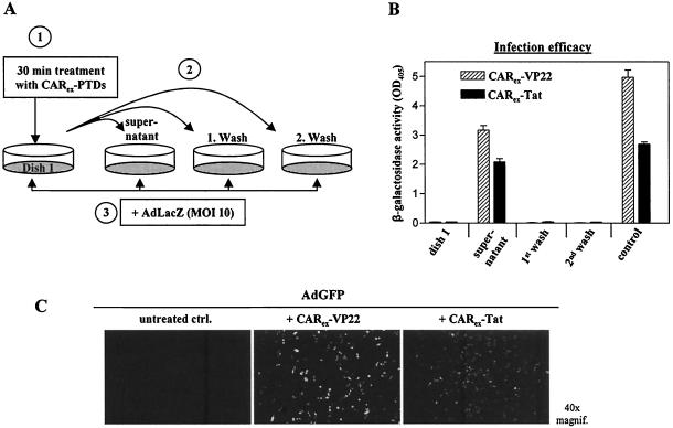
FIG. 6.
CARex-PTD fusion proteins form stable complexes with adenoviral vectors. SKLU-1 cells (dish 1) were treated with CARex fusion proteins (2 nM) and incubated for 30 min. The supernatant and two wash fractions (medium) were then transferred to separate SKLU-1 dishes. For the control sample, the supernatant was left on a dish. Subsequently, all cells were infected with AdLacZ (MOI, 10) for 4 h and incubated for transgene expression. (A) Experimental setup. (B) Results of the corresponding β-galactosidase activity measurements. (C) AdGFP was dialyzed against DMEM and treated with purified recombinant CARex-VP22 or CARex-Tat in a 10-fold molar excess relative to adenoviral fiber knob molecules of AdGFP. As a control, AdGFP received DMEM alone. The virus preparations were then subjected to a CsCl density gradient ultracentrifugation for separation of unbound protein. The virus band was extracted, dialyzed against DMEM, and quantified by determination of optical density at 260 nm. Infection of SKLU-1 cells was carried out for 4 h at an MOI of 50.

