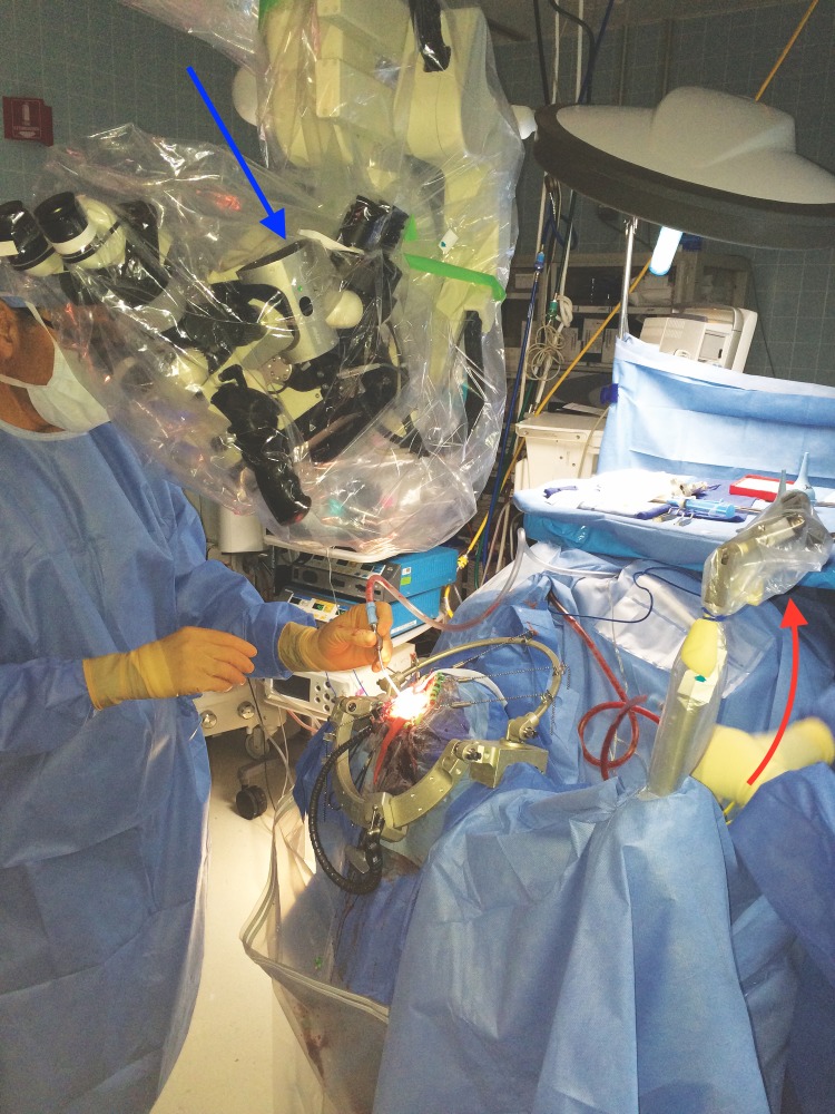
Figure 2. Operative Field.
This figure shows the set up of the operative field. Here, the microscope is fixed in place with the mounted tracker attached (seen at the arrow). Fixing the microscope allowed for the use of the microscope as an extension of the neuronavigational tools. The crosshairs of the microscope are focused on the center of the VBAS retractor, and as the VBAS is advanced, the focus is altered, thereby altering the depth of the tool on the neuronavigation screen. The blue arrow shows the scope-mounted tracker. The red arrow shows the frame-mounted tracker.

