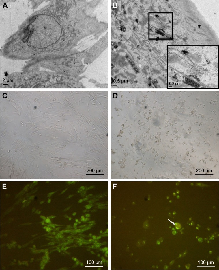
Figure 2.
Cellular uptake of ZnO NPs and morphological changes in treated human MRC5 lung fibroblasts.
Notes: (A) EM micrograph of untreated MRC5 lung fibroblast. (B) EM micrograph of ZnO NP-treated MRC5 lung fibroblast. (C) LM micrograph of untreated MRC5 lung fibroblast. Magnification: ×100. (D) LM micrograph of 50 μg/mL ZnO NP-treated cells. Cells are shrunken, indicative of cell death. Magnification: ×100. (E) Confocal micrograph of untreated MRC5 cell. Magnification: ×200. (F) Confocal micrograph of cells treated with 25 μg/mL ZnO NPs, showing cell shrinkage (as indicated by arrow). Magnification: ×200.
Abbreviations: EM, electron microscope; NPs, nanoparticles; LM, light microscope.

