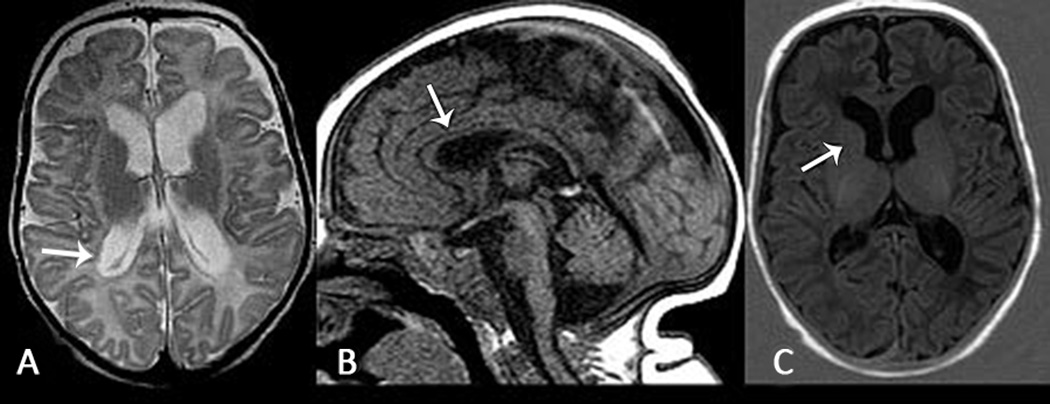
Figure 2. Brain MRI at Three months of age in Infant with KCNQ2 R201C Variant.
A. Axial T2 weighted image shows diffuse reduced brain volume with ex vacuo enlargement of the lateral ventricles, also notice subependymal heterotopias in the right ventricular atrium (white arrow). B. Sagittal T1 weighted image shows diffuse thinning of the corpus callosum (white arrow) that reflects bilateral volume loss in the cortex. C. Axial T1 weighted inversion recovery image shows no myelination in the anterior limb of internal capsule (should be present at three months of age), suggesting hypomyelination (patient A).

