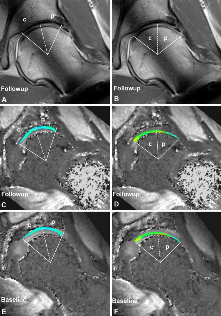
Fig. 4A–F.
The manual placement of regions of interest with help of (A–B) followup morphologic images to define cartilage extension was performed simultaneously on (C–D) followup and (E–F) baseline dGEMRIC images. (A, C, E) The margins for the acetabular cartilage were the rim on the lateral side and the acetabular fossa on the medial side. Cartilage areas were subdivided into (p) peripheral and (c) central regions. (B, D, F) The margins for the femoral cartilage were the head-neck junction on the lateral side and the fovea capitis femoris on the medial side. Cartilage areas were subdivided into (p) peripheral and (c) central regions.

