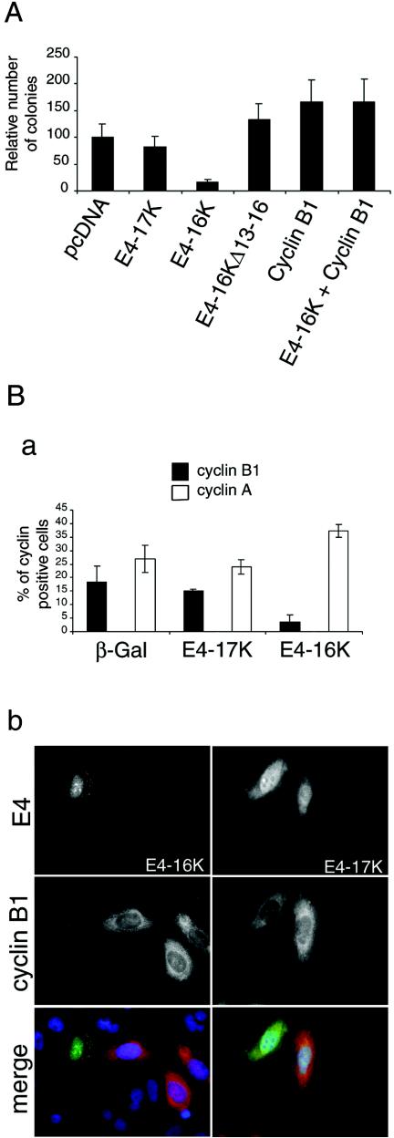
FIG. 6.
Exogenous expression of cyclin B1 rescues E4-16K-induced repression of keratinocyte growth. (A) Graphical representation of the relative number of clones produced following transfection of HeLacells with the different HPV1 E4-containing plasmids, including the G2-defective plasmid E4-16KΔ13-16. Presented data are from three experiments, each with 10 dishes (6 cm) per plasmid. (B) Detection of cyclins A and B1 in SCC-12F cells expressing β-Gal, E4-17K, and E4-16K. Cells infected with the different rAds for 72 h were dual stained for β-galactosidase or E4 and the cyclins A and B1. (a) Histogram of the percentage of dual positive cells. A minimum of 400 cells were counted for each protein, and the results shown are from three independent experiments. (b) Immunofluorescence staining of cells showing examples of cyclin B1-negative E4-16K cells and cyclin B1-positive E4-17K cells. In merged images, E4 proteins are green, cyclin B1 is red, and nuclei are blue. Note that E4-17K is nuclear and cytoplasmic and associated with inclusion bodies, while E4-16K is predominantly in the nucleoplasm and nucleoli (33, 36).

