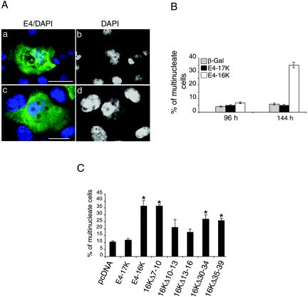
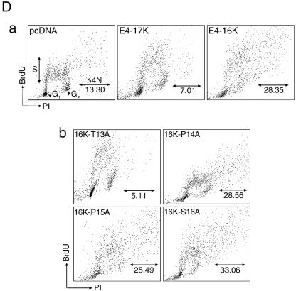
FIG. 7.
E4-16K induces rereplication of the cellular genome. (A) Immunofluorescence microscopy of SCC-12F cells infected with Ad-E416K for 96 h. Cells were stained with an anti-E4 MAb (4.37; green), and nuclei were visualized with DAPI (frames a and c, blue). DAPI staining is also shown in gray scale (b and d). E4-16K-expressing cells contain large and sometimes distorted nuclei (a and b) and are also multinucleate (c and d). Bar, 5 μm. (B) Percentage of β-galactosidase-, E4-17K-, and E4-16K-positive SCC-12F keratinocytes that contain more than one nucleus (up to 5, multinucleate), as determined by immunofluorescence microscopy following infection with rAds. Data were collected every 24 h until 144 h p. i., but only the 96- and 144-h time points are shown. This experiment was repeated in triplicate, and the number of cells counted in each experiment was at least 400. (C) HeLa cells were transfected for 24 h with the empty vector, the E4-containing plasmids, and the panel of E4-16K deletion plasmids. Cells were stained with an E4 MAb, and nuclei were visualized by DAPI staining. The percentage of E4-positive multinucleate cells for each plasmid was recorded; the asterisk indicates a significant (99.9%) increase in multinucleate cells compared to cells transfected with pcDNA. (D) Incorporation of BrdU into DNA of HeLa cells transfected with the empty vector (pcDNA), E4-17K, and E4-16K plasmids (a) and plasmids expressing the alanine point substitutions within 13TPPS16 (b). The distributions of the G1, G2, and S phase populations are indicated. The percentage of BrdU-positive cells in S phase and with a >4N DNA content are as indicated. The G2/M:G1 ratios were 0.61 (pcDNA), 0.64 (E4-17K), 1.10 (E4-16K), 0.55 (16K-T13A), 1.10 (16K-P14A), 0.90 (16K-P15A), and 0.98 (16K-S16A).


