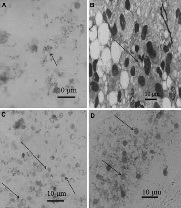
Fig. 1.
Pneumocystis in rat BAL specimen under light microscopy. a Trophozoites of Pneumocystis staining with Geimsa stain (×10), b Cyst walls of Pneumocystis staining with Geimsa stain (×40), c Cyst walls of Pneumocystis staining with Gomori’s methenamine silver stain (×10), d Cyst walls of Pneumocystis staining with Toluidine Blue O stain (×10)

