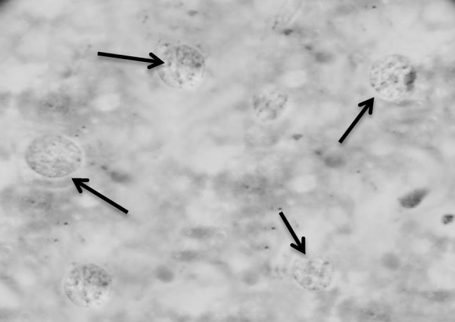
Fig. 11.

Section in murine liver from (G6; SWAP + SEA + adjuvant then infected by S. mansoni) showing hepatocytes with good configuration regarding the state of the nuclei, where the hepatic cell nuclei are vesicular, healthy with intact healthy distributed chromatin, intact nuclear membrane, normal shape and average size, one or two nucleoli and nearly bright red staining of the DNA (black arrows; Feulgen ×1000)
