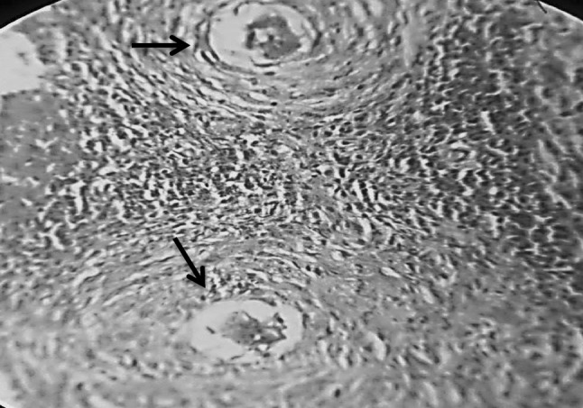
Fig. 2.

Section in murine liver from (G3) (adjuvant then infected by Schistosoma mansoni) showing two close bilharzial fibrocellular granulomas formed of Schistosoma mansoni ova surrounded by lymphocytes, plasma cells, eosinophils, histocytes and little fibrosis (H&E ×400)
