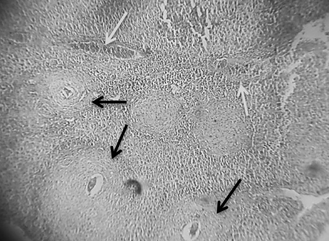
Fig. 4.

Section in murine liver from (G5) (SWAP + adjuvant then infected by S. mansoni) showing moderate sized granulomas (arrows; fibrocellular and fibrous) with central degenerated ova. There was a little bit improvement in the hepatic tissue configuration. Areas of hemorrhages were seen (blue arrow; H&E ×100) (color figure online)
