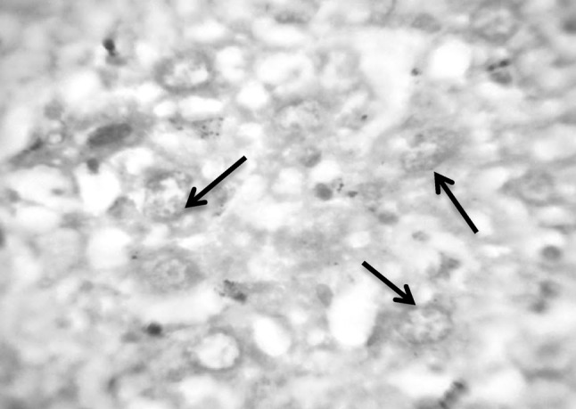
Fig. 7.

Section in murine liver from (G2) control infected mice by S. mansoni: showing hepatocytes in different stages of degeneration with vacuolated cytoplasm. The nuclei showed fragmentation (arrows), chromatolysis, partially destroyed nuclear membrane and very pale red staining of the DNA (Feulgen ×1000)
