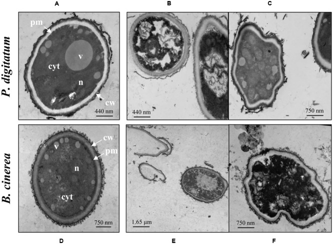
FIGURE 3.

Effect of harmol on ultrastructure of conidia. Transmission electron micrographs of the indicated pathogens: (A,D) conidia in control treatment; (B,C,E,F) conidia treated with 1 mM harmol. Photographs are representative of three independent experiments. cyt, cytoplasm; v, vacuole; n, nucleus; pm, plasmatic membrane; cw, cell wall.
