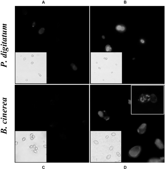
FIGURE 4.

Effect of harmol on integrity of the cell wall. Conidia were exposed to 1 mM harmol during 24 h and incubated with CFW. Fluorescence microscopy images (100×) of conidia in control treatment (A,C), and treated conidia (B,D). The corresponding light microscopy images are shown for each panel. Photographs are representative of three independent experiments.
