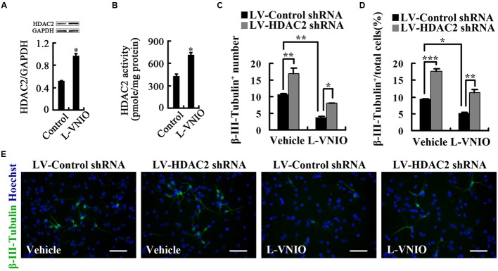
FIGURE 6.
Repression of nNOS negatively regulates differentiation of embryonic NSCs into neurons by upregulation of HDAC2. (A,B) HDAC2 levels (A) and enzymatic activity (B) in cultures treated with 100 μM L-VNIO or vehicle for the first 24 h during differentiation. (C–E) HDAC2 down-regulation rescues L-VNIO-induced neuronal differentiation reduction. 100 μM L-VNIO or vehicle was treated for the later 2 days during the 4-day differentiation of LV-HDAC2 shRNA- or LV-Control shRNA-infected NSCs. (C) Bar graph showing the number of newborn neurons. (D) Bar graph showing the percentage of newborn neurons. (E) Representatives of β-III-Tubulin+ neurons. Scale bars = 50 μm. Data are mean ± SEM (n = 3); ∗p < 0.05, ∗∗p < 0.01, ∗∗∗p < 0.001. GAPDH, glyceraldehyde phosphate dehydrogenase; HDAC2, histone deacetylase 2; LV-Control shRNA, lentiviral vector containing control shRNA; LV-HDAC2 shRNA, lentiviral vector containing shRNA of HDAC2; L-VNIO, N5-(1-imino-3-butenyl)-L-ornithine.

