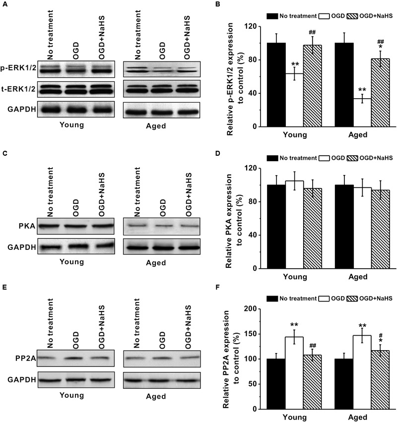
FIGURE 6.

Involvement of ERK pathway in the neuroprotection of NaHS. (A,B) OGD treatment inhibited p-ERK1/2 (MW: p-ERK1/2, 44/42 kDa; EKR1/2, 44/42 kDa) expression in both young and aged hippocampal neurons, while 250 μM NaHS could increase ERK1/2 phosphorylation, characterized by western blot analysis. (C,D) Western blotting shows that there was no significant difference in PKA (MW: 45 kDa) levels between the experimental groups. (E,F) OGD increased PP2A (MW: 35 kDa) expressions, while NaHS decreased its expressions. The expression levels from each group were normalized to those of GAPDH (as a loading control, MW: 37 kDa) and are presented as ratios to control. Data were presented by mean ± SD. ∗p < 0.05 and ∗∗p < 0.01 versus corresponding control (no treatment), ##p < 0.01 versus corresponding OGD group.
