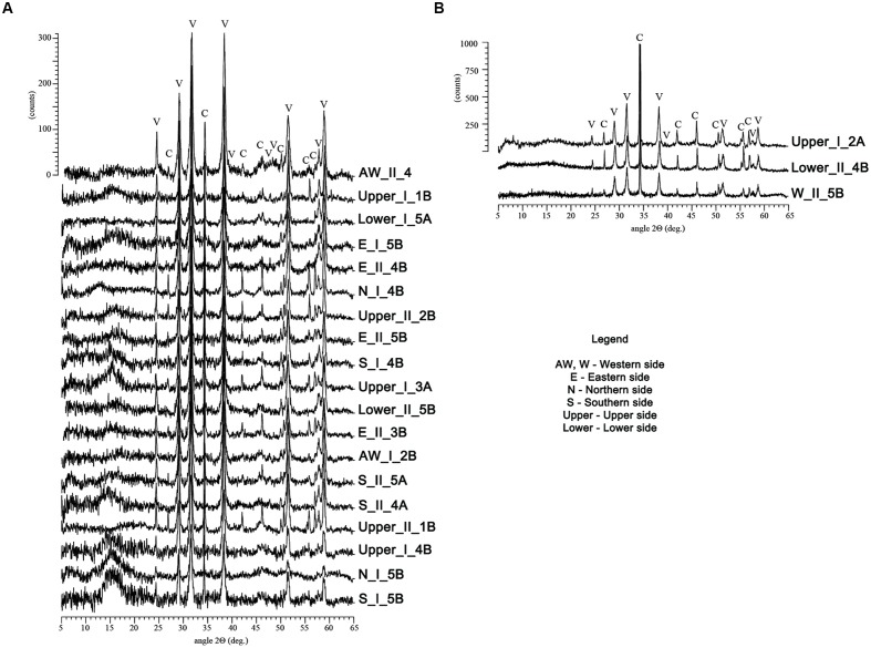
FIGURE 4.
X-ray diffraction patterns of the crystals recovered from the M-3 agar media recorded on a Bruker D8 Advance diffractometer. The patterns of the samples in the (A) image were predominated by vaterite (V), while the ones from the (B) contained predominantly calcite (C). The y-scale (intensity) was fairly similar for each group of samples.

