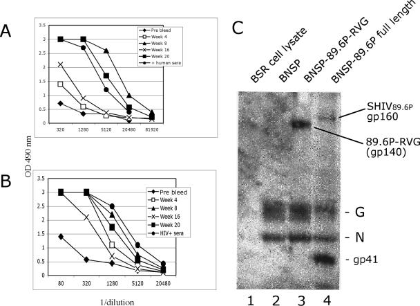
FIG. 1.
Immune responses after vaccination. (A) Anti-RV RNP response after vaccination with ΔG-89.6P-RVG. The animal received intramuscular, i.v., and subcutaneous inoculations at day 0 and week 7 and an additional i.v. booster on week 19. The positive control is human serum from an RV-vaccinated individual. OD 490 nm, optical density at 490 nm. (B) Anti-HIV-189.6 Env response in a vaccinated animal. Oligomeric HIV-189.6 gp140 was coated onto a microtiter plate and reacted with serial dilutions of rhesus serum from the indicated time points as indicated. Serum from an individual chronically infected with HIV-1 served as a positive control. (C) Western blot analysis of week 8 serum from an immunized macaque. Cell lysates were prepared from uninfected BSR cells (lane 1), cells infected with an empty RV vector (lane 2), an RV vector expressing SHIV89.6P Env containing the cytoplasmic domain of RV G (lane 3), or RV vector expressing SHIV89.6P Env containing the entire gp160 coding region (lane 4).

