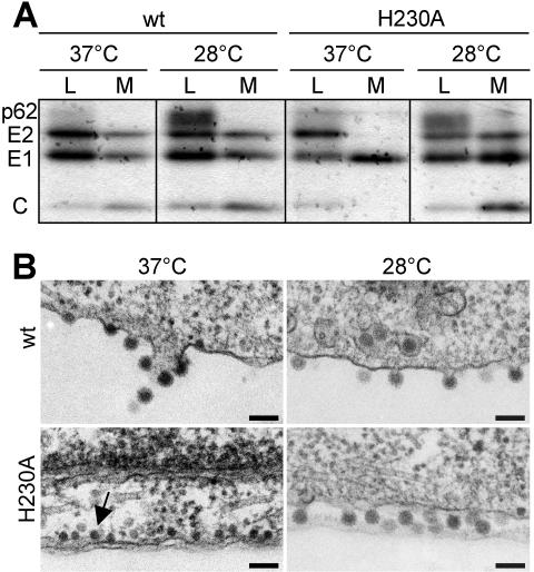
FIG. 2.
Assembly of wt SFV and H230A mutant in BHK cells. BHK cells were electroporated with wt-ic or H230A-ic RNA and incubated at 37°C for 2 h. Cells were further incubated for 4 h at 37°C or overnight at 28°C before analysis as described. (A) Protein expression and virus assembly. Cells were pulse-labeled with [35S]methionine and cysteine and chased at 37 or 28°C. SFV proteins in the cell lysates (L) and medium (M) were immunoprecipitated with a polyclonal antibody to E1 and E2, and samples were analyzed by SDS-PAGE. Chase medium samples were immunoprecipitated in the absence of detergent to allow recovery of intact virus particles containing the viral nucleocapsid. The position of the different viral proteins is indicated (C, capsid). Data shown are representative of three experiments. (B) Electron microscopy of wt or H230A virus-infected-cells. The arrow indicates the presence of viral nucleocapsids underneath the plasma membrane. Bar, 0.1 μm.

