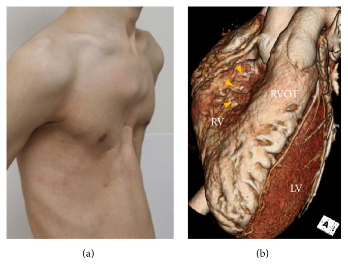
Figure 1.

(a) Gross picture shows a deformity of the anterior thoracic cage consistent with pectus excavatum. (b) 3D reconstructed computed tomography image also reveals external compression of basal-to-mid portion of the right ventricle (yellow arrow heads). LV, left ventricle; RV, right ventricle; RVOT, right ventricular outflow tract.
