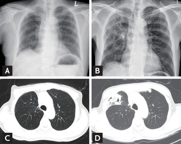
Figure 1.

(A) Simple chest X-ray before tuberculosis (TB) reactivation. There is no cavitary lesion. (B) Simple chest X-ray at the time of TB reactivation diagnosis. There are cavitary lesions on the lung right middle lobe. (C) High resolution computed tomography (HRCT) before TB reactivation. (D) HRCT at the time of TB reactivation diagnosis.
