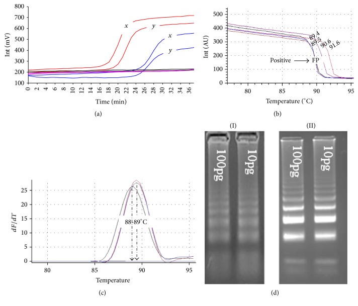
Figure 2.
(a) The tube scanner fluorometric output of stem LAMP (red) and standard LAMP (blue) tests using ~10 pg of DNA from isolate PT41 (x) and DNA prepared from CSF (y) of a confirmed HAT patient. The unit reports the fluorescence in millivolts (mV) on y-axis and time in minutes on the x-axis. (b) The acquisition of melts curves for PT41, DNA prepared from CSF, and supernatant prepared from a tsetse fly sample spiked with B014 DNA. Tm was 89.5°C for PT41 and DNA from CSF patient and 89.4°C for B014 in tsetse fly sample. The nonspecific product (FP) is induced for illustration and showed Tm that ranged from 72 to 76°C and 90 to 92°C (c). The duplication of melts peaks for PT41, CSF, and B014 DNA samples using Rotorgene 6000. The melt peaks showed Tm of ~89°C for PT41 and CSF DNA and ~ 88°C for B014. (d) The amplicons for stem LAMP test (I) without outer primers and (II) with outer primers.

