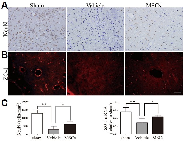
Fig. 3. Neural protection and alterations to tight junction expression after treatment.

(A) Transplantation of MSCs increased the number of NeuN+ cells in the perihematomal region. (B) Brain sections from the sham group, HICH-vehicle group and HICH-MSC group were analyzed for ZO-1 by a fluorescence microscope at 3 days following HICH. Increased intensity of ZO-1 expression was observed in the HICH-MSC group compared with the HICH-vehicle group. (C) Quantification of cell marker expression showed a significant increase in the number of NeuN+ cells (n = 4) and the mRNA expression levels of ZO-1 in the HICH-MSC group at 3 days (n = 5), compared with the HICH-vehicle group. ** p < 0.01; * p < 0.05. Scale bar: 50 μm.
