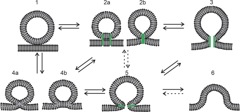
Figure 1.
Putative intermediates of membrane fusion and their transitions. Transitions that are less relevant or speculative are indicated by dashed arrows. Protein elements are colored green. (1) A vesicle approaches the plasma membrane. (2) Proteins hold the vesicle and plasma membrane together, either through separate contacts (a) or through one central contact (b). (3) A proteinaceous fusion pore could form from a central contact as in 2b. (4) Lipid mixing of the outer (proximal) leaflets can begin, first through the formation of a stalk (4a) and then through the formation of an extended hemifusion diaphragm (4b) in which the fused proximal leaflets retract and leave a bilayer formed by the two distal leaflets. (5) A fusion pore formed by a contiguous lipid bilayer curved into an hourglass-like shape. (6) A greatly expanded lipid fusion pore on the way to complete merger of the plasma and vesicle membranes.

