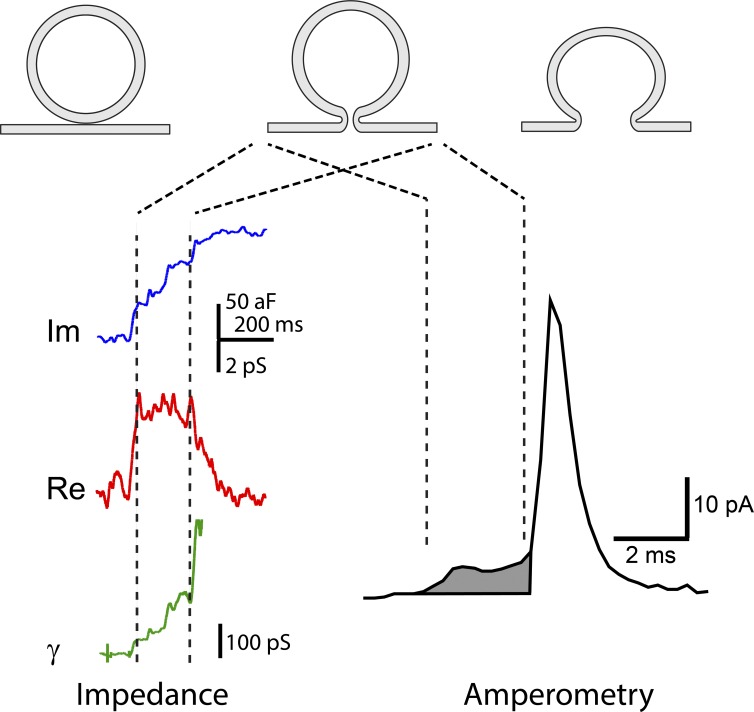
Figure 3.
Impedance and amperometry measurements of fusion pores can be interpreted in terms of three successive stages of membrane fusion. (1) Contact (top left), (2) fusion pore opening (top middle), and (3) fusion pore expansion (top right). (left) Impedance recording reveals fusion pore openings as a change in the complex impedance of a patch of membrane to which a vesicle fuses. The imaginary component of the impedance (blue) and real component (red) are used to calculate the fusion pore conductance, γ (green; Lollike et al., 1995). The opening of a fusion pore produces an initial conductance increase, and fusion pore expansion increases the conductance further. (right) Amperometry recording reveals a fusion pore opening as a pre-spike foot (shaded), which represents the flux of catecholamine out of the vesicle at a limited rate. Fusion pore expansion allows content release much more rapidly to produce an amperometric spike.

