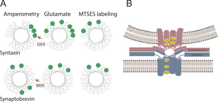
Figure 4.
SNARE TMD residues lining the fusion pore. (A) Residues in the SNARE TMDs identified by various fusion pore measurements are highlighted in green. The first TMD residue of the wheel is labeled with a red arrow. Amperometry identified some residues (Han et al., 2004; Chang et al., 2015). Glutamate flux and labeling by MTSES identified some of the same residues as well as some different residues (Bao et al., 2016). Many of the residues identified in a given assay fell along one face of a helical wheel (wheels generated at http://kael.net/helical.htm). (B) Fusion pore model based on TMD mutagenesis and amperometric pre-spike feet and conductance in endocrine release (Chang et al., 2015).

