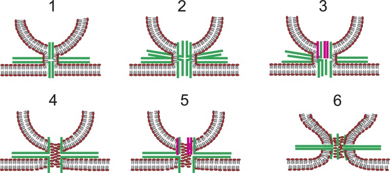
Figure 9.
Composite lipid–protein fusion pores. SNAREs are green; synaptophysin TMDs are pink. Models 1–3 illustrate continuity of the proximal monolayer of the vesicle and plasma membrane outside a proteinaceous fusion pore. These models have no contact between phospholipids and the aqueous pore lumen. Models 4–5 illustrate lipid headgroups of a bilayer that forms a pore in which lipid and protein alternate along the walls. Model 6 illustrates protein TMDs lodged among the headgroups of a lipidic fusion pore.

