Figure 1.
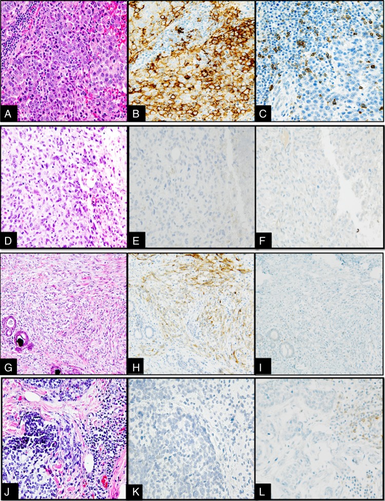
(A–L) Interface between tumour and tumour infiltrating lymphocytes (TILs) in different metaplastic breast carcinomas (MBCs) categorised into four categories based on programmed death-ligand 1 (PD-L1) and programmed cell death 1 (PD-1) expression, 400× magnification. (A–C) Type 1 (PD-L1 positive, high PD-1): MBC with squamous metaplastic component (A) showing 3+ intensity PD-L1 staining in 50% of the tumour (B) and high PD-1 expression in the peritumoral lymphocytes (210/10 high power fields) (C). (D–F) Type 2 (PD-L1 negative, low PD-1): MBC with spindle cell metaplastic component (D) with tumour cells showing no increase in expression of PD-L1 by tumour cells (E) and no expression of PD-1 by interstitial lymphocytes/plasma cells (F). (G–I) Type 3 (PD-L1 positive, low PD-1): MBC with spindle cell metaplastic component (G) with moderate overexpression of PD-L1 in the tumour cells (H) and no expression of PD-1 in the TILs (I). (J–L) Type 4 (PD-L1 negative, high PD-1): MBC with areas of chondroid metaplastic component (J) with no PD-L1 overexpression in tumour cells (K) and moderate expression of PD-1 positive in TILs (190/10 high power fields) (L).

