
Figure 2.
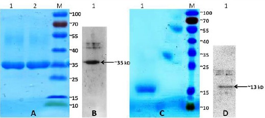
Purification and expression of the recombinant CFP-10:Fcγ2a and CFP-10:His fusion proteins. A) Analysis of the recombinant CFP-10:Fcγ2a protein by 12% SDS-PAGE stained with Coomassie Blue. Lane 1, 2: fractions of the eluted recombinant protein of approximately 35 kDa. Lane M: protein marker. B) The expression of the recombinant CFP-10:Fcγ2a protein was analyzed by western blot using anti mouse IgG-HRP. C) Analysis of the recombinant CFP-10:His protein by 12% SDS-PAGE stained with Coomassie Blue. Lane 1: fractions of the eluted recombinant protein of approximately 13 kDa. Lane M: protein marker. D) The expression of the recombinant CFP-10:His protein was analyzed by Western blot using anti His tag-HRP. Lane M: protein marker