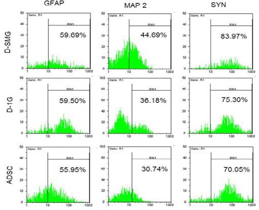
Figure 8.

After ADSCs were cultured for 24 hr in differentiation medium, flow cytometry analysis of neural markers (GFAP, MAP-2, and synaptophysin) were done from ADSCs (control), D- SMG(differentiation in SMG) groups, and D-1G (differentiation in 1G) group
