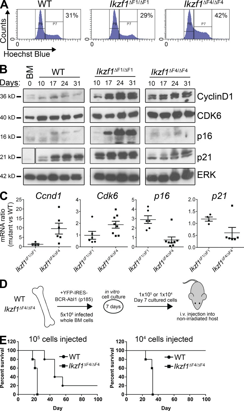
Figure 1.
Loss of Ikaros tumor suppression in Ikzf1ΔF4/ΔF4 mutant mice results in an increase in active cell cycle and aggressive leukemia in a nonirradiated in vivo model. (A–C) BM from WT, Ikzf1ΔF1/ΔF1, and Ikzf1ΔF4/ΔF4 mutant mice were transduced with BCR-ABL1-p185-IRES-YFP and grown in vitro on BM stroma–derived feeder layers. (A) Cell cycle flow cytometry analysis was performed by Hoechst incorporation, and one representative experiment (at day 14 of cell culture) is shown. (B) Cells were harvested on different days of in vitro cell culture and protein was extracted for Western blot analysis of Cyclin D1, Cdk6, p16, and p21, with ERK as loading control. Space indicates that intervening lanes have been spliced out. (C) RNA was isolated from the in vitro cultures, cDNA was prepared, and mRNA for Ccnd1, Cdk6, p16, and p21 was analyzed by quantitative real-time PCR. Ubc was used as a reference gene. (D) Schematic of the experimental set up for the in vivo transplantation assay. BM cells from WT and Ikzf1ΔF4/ΔF4 mutant mice were transduced and cultured in vitro as in A–C for 7 d before i.v. injection into nonirradiated immunocompetent WT c57BL/6 recipient mice. Animals were monitored and euthanized upon signs of leukemia development. (E) Kaplan-Meyer curve of cohorts receiving 105 (left) or 104 (right) BCR-ABL1–transformed cells. (A–C) are representative of three independent experiments and (E) represents one experiment with five recipient mice for each of the four cohorts. The result in right panel was repeated in a separate independent experiment displayed in Fig. 6 F.

