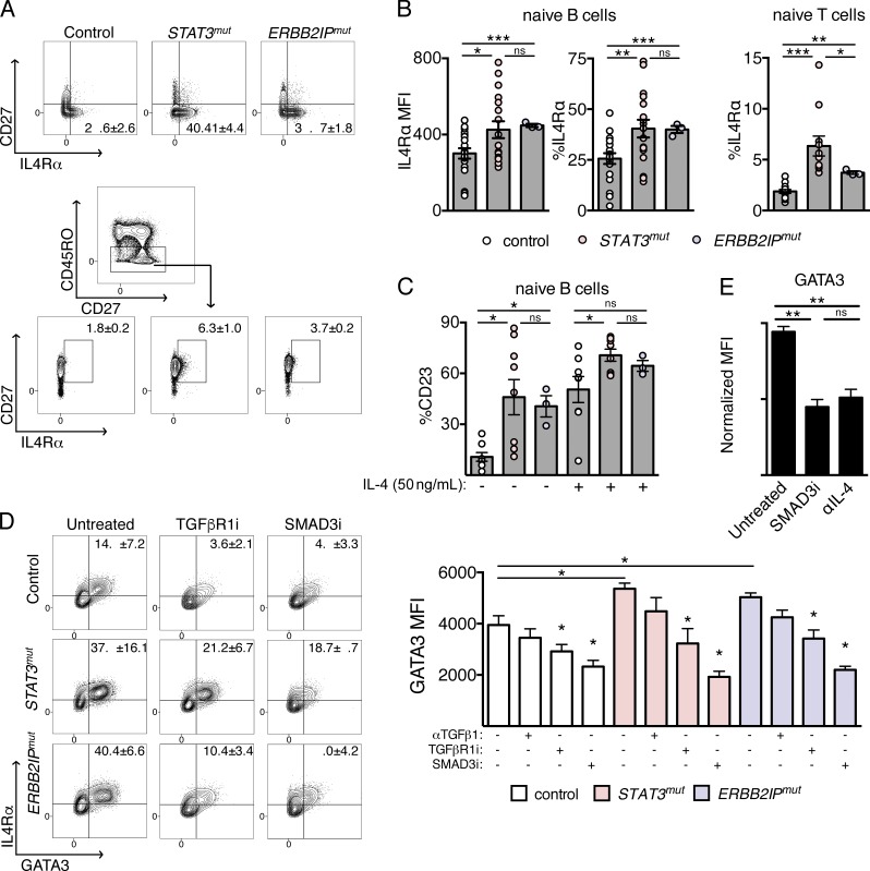
Figure 5.
Increased functional IL-4Rα expression in STAT3mut and ERBB2IPmut naive lymphocytes promotes TGF-β–dependent GATA3 expression in CD4 T cells. (A) Contour plots demonstrating expression of IL-4Rα ex vivo on naive (CD27−) B cells (top) and contour plots showing CD27 and IL-4Rα coexpression and gating strategy of naive (CD45RO−) T cells (bottom) from control, STAT3mut, and ERBB2IPmut patients. (B) Combined ex vivo IL-4Rα expression on naive lymphocytes from control (n = 19), STAT3mut (n = 17), and ERBB2IPmut (n = 3) individuals. MFI on naive (CD27−) B cells (left) and percentage of naive (CD27−) B cells (middle) and of CD27+ naive (CD45RO−) CD4 cells (right) expressing IL-4Rα are shown. (C) Percentage of naive (CD27−) B cells from controls (n = 8), STAT3mut (n = 8), and ERBB2IPmut (n = 3) individuals expressing the STAT6-target CD23 ex vivo or after short-term stimulation with IL-4 as indicated. (D) IL-4Rα and resulting GATA3 expression in control, STAT3mut, and ERBB2IPmut naive CD4 T cells after activation (CD69+) with weak TCR stimulation under nonskewing conditions in the presence or absence of a TGF-β1–neutralizing antibody (αTGFβ1), a TGFβR1 inhibitor (TGFβR1i), or a selective SMAD3 inhibitor (SMAD3i). Contour plots (left) and combined GATA3 MFI (right) are shown. (E) Comparison of GATA3 expression in activated (CD69+) control naive CD4 cells, as normalized MFI, cultured with 10 µg/ml neutralizing IL-4 antibody (αIL-4) or SMAD3i under the same conditions as in D. Data are representative or combined from at least three independent experiments and are represented as the mean ± SEM. Unpaired two-tailed Student’s t tests and Mann-Whitney tests were used where appropriate. *, P < 0.05; **, P < 0.005; ***, P < 0.001.

