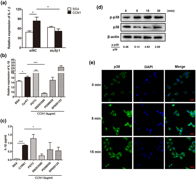
Figure 4. The α6β1/p38 MAPK pathway was activated in keratinocytes.
(a) IL-1β mRNA in CCN1-administrated HaCaT cells treated with specific α6β1 siRNA. (b) The mRNA level of IL-1β in CCN1-challenged keratinocytes. (c) The protein level of IL-1β in CCN1-stimulated keratinocytes. For (b,c), keratinocytes were treated with 4 μM PDTC, 10 μM SB203580, 1 μM PD98059, or 20 μM SP600125 in combination with CCN1 (5 μg/ml) (shadow bars) for 2 h. Open bar, BSA, black bar, CCN1 with no inhibitors. (d) Phosphorylation of p38 in CCN1-stimulated HaCaT detected by western blotting analysis. (e) Nuclear translocation of p38 in CCN1-stimulated HaCaT cells monitored by immunofluorescence. p38 was detected by FITC-anti-p38 (green). Nuclei were stained with DAPI (4,6-diamidino-2-phenylindole; blue). This merged picture shows p38 translocation to the nucleus. Bar, 20 μm. CCN1 at 5 μg/ml was used to stimulate keratinocytes. *P < 0.05, ***P < 0.005. The data represent one of three independent experiments.

