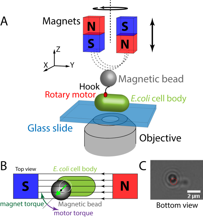
Figure 1. Magnetic tweezers assay for studying the E. coli flagellar motor.
(A) Schematic of the experimental configuration (not to scale). The magnetic bead (grey) is connected to the flagellar motor (red) of E. coli by its flexible hook (black). The E. coli cell body (green) is fixed to a glass slide (blue). Two permanent magnets generate the external magnetic field (dashed grey lines). The objective images the sample plane onto a camera. (B) Top view of the experimental configuration (not to scale). The motor rotates the bead counter clockwise (purple arrow, the motor torque vector points out of the plane), while the magnet torque tends to align the magnetic bead with the field lines. At this orientation of the bead, the magnet torque vector points into the plane and pull the bead in the clockwise direction. (C) Camera image (bottom view) of a magnetic bead (black circle with white centre and diffraction rings) rotated by an E. coli cell (scale bar 2 μm). The bead traces out a circle indicated by the red dashed line.

