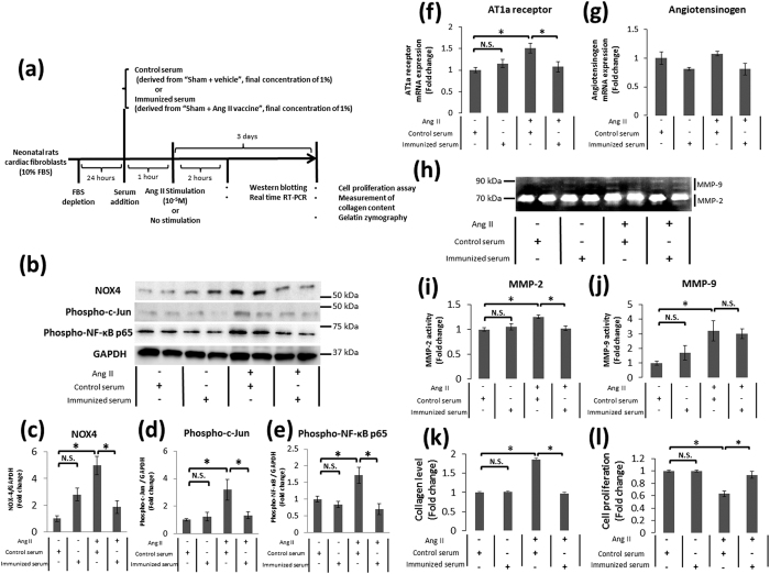
Figure 7. The potency of the antibody produced by the Ang II vaccine on post-MI remodeling-associated responses in cardiac fibroblasts.
(a) The protocol to evaluate the potency of immunized serum addition on Ang II signaling. (b) Representative Western blots of NADPH oxidase 4 (NOX4), phospho-c-Jun, phospho-nuclear factor-κB p65 subunit (NF-κB p65), and glyceraldehyde 3-phosphate dehydrogenase (GAPDH) in neonatal rat cardiac fibroblasts. The relative protein expression of (c) NOX4, n = 4 each; (d) phospho-c-Jun, n-4 each; and (e) phospho-NF-κB p65, n = 5-6 each. *p < 0.05 by the Tukey–Kramer post hoc test. The relative mRNA expression of (f) angiotensin type 1a receptor (AT1aR), n = 5-6 each; and (g) angiotensinogen, n = 5–6 each. *p < 0.05 by the Tukey–Kramer post hoc test. (h) A representative image of gelatin zymography for matrix metalloproteinase (MMP)-2 and MMP-9 in the conditioned media of cultured cardiac fibroblasts. The relative activity of (i) MMP-2, n = 6 each; and (j) MMP-9, n = 6 each. *p < 0.05 by the Tukey–Kramer post hoc test. (k) The relative collagen level in cardiac fibroblasts, n = 12 each; and (l) the proliferation ratio of cardiac fibroblasts, n = 12 each. *p < 0.05 by the Tukey–Kramer post hoc test.

