
FIGURE 7.
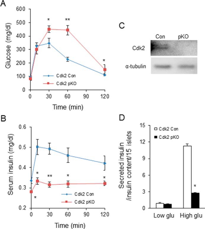
β-Cell dysfunction upon CDK2 deletion. Shown are GTT (A) and serum insulin levels (B) during the GTT in 2-month-old male Cdk2-Con (diamonds) and Cdk2-pKO (squares) mice (n = 7 in each genotype). C, Western blotting analyses to compare CDK2 protein expression in islets from Cdk2-WT (Con) and Cdk2-pKO mice. α-Tubulin protein levels are shown as loading control. D, glucose-stimulated insulin secretion under low (3.3 mm) and high (25 mm) glucose condition in Cdk2-Con (open bars) and Cdk2-pKO (closed bars) 2-month-old male mice islets (n = 15 islets/pancreas harvested from five mice of each genotype). Insulin secretion is normalized by total cellular insulin content of islets. The data comprises results derived from three independent experiments. Statistical analysis was performed with Student's t test; *, p < 0.05; **, p < 0.001. Error bars, S.D.