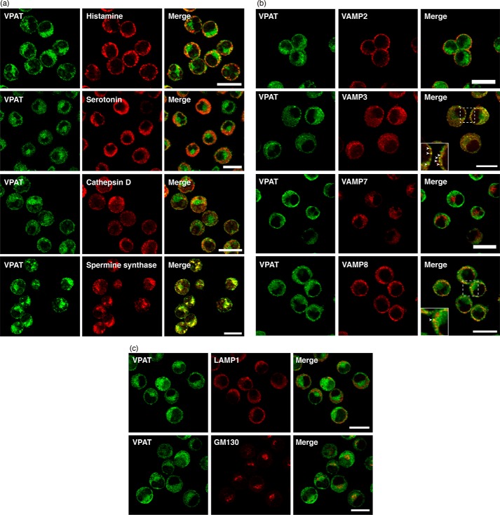
FIGURE 2.
Localization of VPAT in BMMCs. a–c, BMMCs were fixed and subjected to double immunostaining with antibodies to VPAT (green, left), histamine, serotonin, cathepsin D, and spermine synthase (a); VAMP2, VAMP3, VAMP7, and VAMP8 (b); and LAMP1 and GM130 (c) (red, middle). Merged images (right) are also shown. Areas surrounded by dotted lines are enlarged in insets. Arrowheads, merged regions. Bars, 10 μm.

