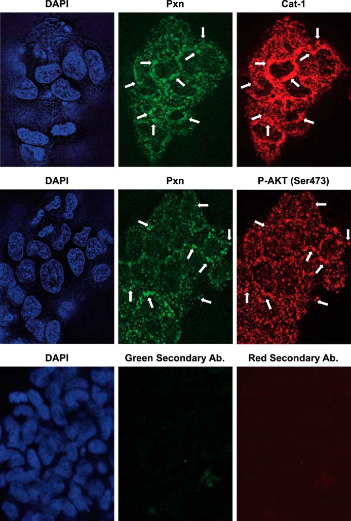
FIGURE 4.

Paxillin, Cat-1, and activated AKT localize together in HeLa cells grown under anchorage-independent conditions as spheres. HeLa cells were cultured under anchorage-independent conditions by plating them on bacteria-grade dishes and allowing them to form spheres in suspension. The spheres were then collected and stained with the indicated combinations of Pxn, Cat-1, and phospho-Akt (Ser-473) antibodies. As a negative control, spheres of HeLa cells were also stained with only the fluorescently conjugated secondary antibodies (Green Secondary Ab and Red Secondary Ab). DAPI was used to label nuclei. Arrows indicate some of the areas where paxillin localizes with either Cat-1 or phospho-Akt (Ser-473). The experiments shown were performed three separate times, each yielding similar results.
