Figure 1.
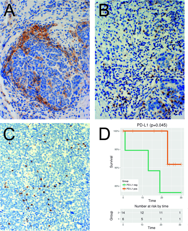
(A) Immunohistochemistry (IHC) of PDL1, 100×: Tumor cells stain positively for PDL1 mainly at the tumor-stroma border. (B) IHC of PD1, 100×: At the tumor-stroma border clusters of immune cells and TILs with PD1 positive cytoplasmatic +/− membrane staining were observed. (C) IHC of CD8, 100×: CD8 positive TILs infiltrating the tumor. (D) The positive impact of PD-L1 expression (≥1% versus 0%) on overall survival (months) shown by a Kaplan-Meier estimate (y-axis is truncated to see the difference more pronounced; p = 0.045; Log-rank test).

