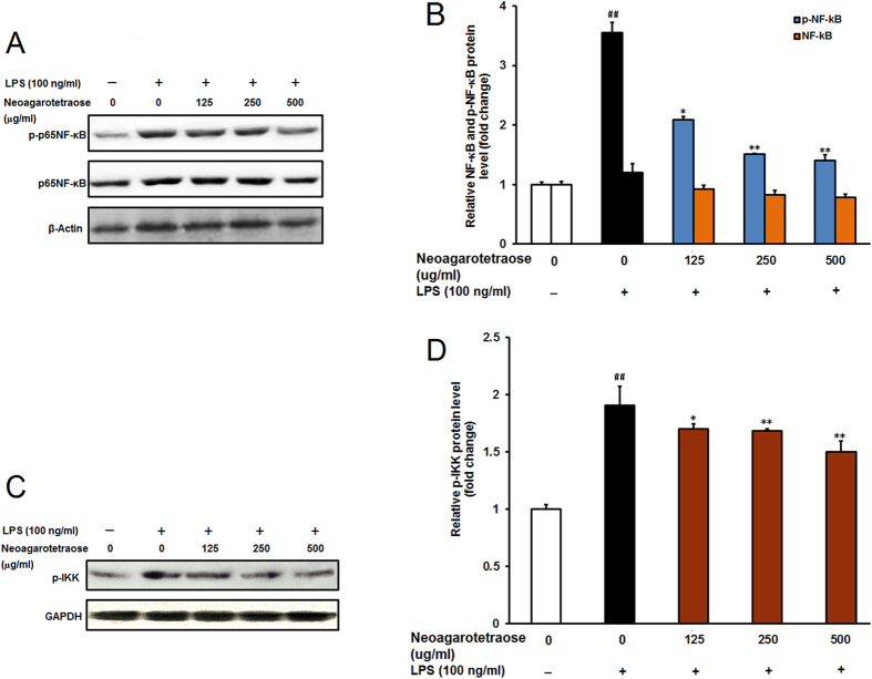
Figure 6. Effect of neoagarotetraose on the activation of NF-κB pathway in RAW264.7 cells.
(A) After pretreated with neoagarotetraose (125, 250, 500 μg/ml) for 2 h, the LPS (100 ng) was added to cells and incubated for 1 h. Then the expression levels of phosphorylated NF-κB and total NF-κB were detected by western blot analysis, respectively. Blots were also probed for β-actin as loading controls. The result shown is a representative of three separate experiments with similar results. (B) Quantification of immunoblot for the ratio of total NF-κB and phosphorylated NF-κB to β-actin. The ratio for non-treated normal control cells was assigned a value of 1.0 and the data presented as mean ± SD (n = 3). Significance: ##P < 0.01 vs. normal control; *P < 0.05, **P < 0.01 vs. LPS treated control. (C) After pretreated with neoagarotetraose (125, 250, 500 μg/ml) for 2 h, the LPS (100 ng) was added to cells and incubated for 1 h. Then the levels of phosphorylated IKK were detected by western blot. Blots were also probed for GAPDH as loading controls. (D) Quantification of immunoblot for the ratio of phosphorylated IKK to GAPDH. The ratio for non-treated normal control cells was assigned a value of 1.0 and the data presented as mean ± SD (n = 3). Significance: ##P < 0.01 vs. normal control; *P < 0.05, **P < 0.01 vs. LPS treated control.

