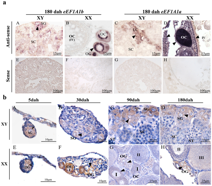
Figure 2. Cellular localization of eEF1A1b in tilapia testis and ovary at different developmental stages by ISH and IHC.
(a) Cell type expressing eEF1A1a and eEF1A1b in tilapia gonads by ISH. eEF1A1b was detected in spermatogonia of the testis, while in the oogonia and phase I oocytes of the ovary at (A and B). eEF1A1a was detected in somatic cell of the testis, while in the oocytes and somatic cells of the ovary (C and D). (b) Cell type expressing eEF1A1b in tilapia gonads by IHC. Consistent with in situ hybridization results, eEF1A1b was detected in the spermatogonia of the testis from 5 to 180 dah (A–D), while in the oogonia of ovary at 5 dah, later in the oogonia and phase I oocytes of the ovary from 30 to 180 dah (E-H). SG, spermatogonia; SC, spermatocytes; ST, spermatids; OG, oogonia; OC, oocytes; I-IV, phase I to phase IV oocytes; Arrowheads indicate the positive signal.

