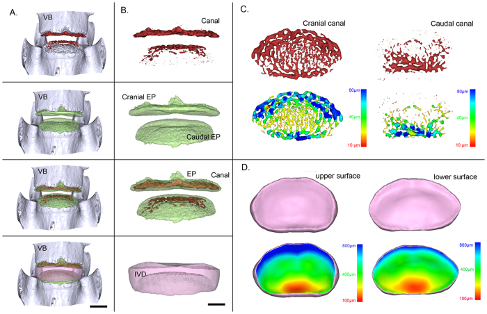
Figure 3. Representative 3D images of the canals in the endplate and IVD and corresponding pseudo-color images obtained by PPCST.
(A) Intact 3D morphology of the lumbar functional unit. (B) 3D images of the canals, endplate, canals located in endplate and IVD. (C) Original and pseudo-colored images of the 3D canal network in the cranial and caudal endplates. (D) Original and pseudo-colored images of the upper and lower surfaces of the IVD. The pseudo-color bar in the lower-right corner indicates the diameter ranges of the canals in the endplate or the IVD thickness distribution.

