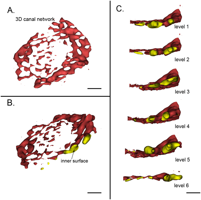
Figure 4. Virtual micro-endoscopy of the canals in the endplate.
(A) Lateral view of the 3D canal network extracted from the endplate. (B) The inner surface of the 3D canal network is clearly present. (C) Virtual navigation within the multiple levels of the internal surface of the canal. (A) and (B) scale bar = 1 mm, (C) scale bar = 500 μm.

