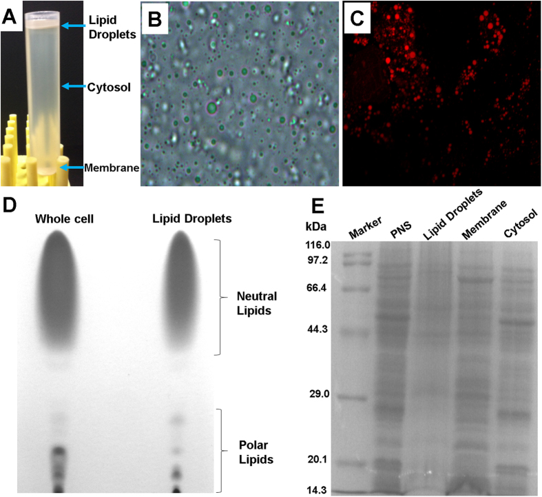
Figure 3.
(A) Naked-eye appearance of a centrifuged post-nuclear supernatant (PNS) sample; (B) Light microscopy image of the isolated LDs; (C) Fluorescent microscope image of the isolated LDs; (D) Thin-layer chromatography of lipid samples from whole cells and LDs; (E) SDS-PAGE analysis of proteins from the PNS, LD, membrane, and cytosol fractions.

