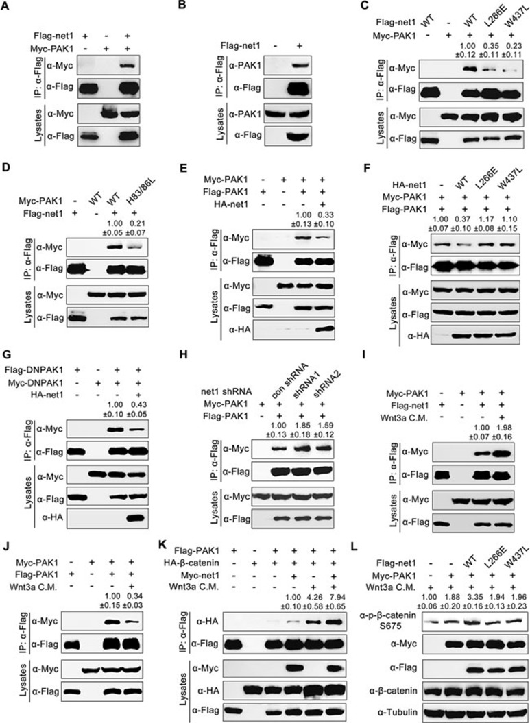
Figure 6.
Effect of Net1 on PAK1 dimerization. (A-D) Flag-tagged Net1 interacts with overexpressed (A) or endogenous (B) PAK1. HEK293T cells were transfected as indicated with expression plasmids encoding Myc-tagged PAK1 and Flag-tagged Net1 or Net1 mutants, then harvested for immunoprecipitation with anti-Flag M2 agarose beads. Note that compared with the wild-type control, Flag-Net1-L266E or W437L has a much lower binding affinity for Myc-PAK1 (C). Conversely, Myc-tagged PAK1 H83L/H86L mutant (which cannot bind Rho family small G proteins such as Cdc42 and Rac1) loses the ability to associate with wild-type Net1 (D). (E and F) Wild-type Net1 (E) but not its L266E or W437L mutant (F) disrupts dimerization of PAK1. HEK293T cells were co-transfected with plasmids expressing Flag- or Myc-tagged PAK1 and HA-tagged wild-type Net1 or Net1 mutants as indicated. Lysates were immunoprecipitated with anti-Flag M2 agarose beads and immunoblotted as indicated. (G) Net1 disrupts the dimerization of DNPAK1, the kinase-inactive mutant. HEK293T cells were co-transfected with plasmids expressing Flag- or Myc-tagged PAK1 K299A mutant proteins and HA-tagged Net1. Lysates were subjected to immunoprecipitations and immunoblots. (H) net1-depletion enhances PAK1 dimerization. HEK293T cells transfected with net1 shRNA1 or shRNA2 were harvested for immunoprecipitation and immunoblotting. (I and J) Wnt3a CM treatment enhances the interaction between PAK1 and Net1, and disassociates the dimerization of PAK1. HEK293T cells co-transfected with indicated plasmids were treated with or without Wnt3a C.M. for 12 h before harvest, and then lysed for immunoprecipitations. (K and L) Net1 enhances the binding affinity of PAK1 for β-catenin (K) and promotes PAK1-induced phosphorylation of β-catenin S675 (L) in the presence of Wnt3a. HEK293T cells transfected with the indicated constructs were treated with or without Wnt3a CM for 12 h before harvesting for immunoprecipitation and western blot analyses. In C-K, quantification is the mean relative ratio of co-immunoprecipitated signals over lysate, while in L, quantification is the relative density of phospho-specific signals to corresponding total protein signals (mean ± SD, three independent biological repeats).

