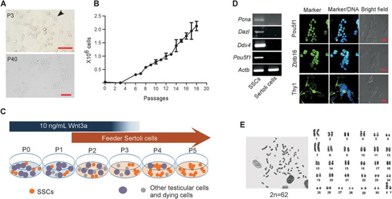
Figure 3.
Establishment of tree shrew spermatogonial stem cell lines. (A) Thy1+ cells were cultured in the presence of 10 ng/mL Wnt3a and Sertoli cell feeder. Images show the appearance of germ cell clumps at passage 3 (P3, upper panel, arrow head), and stem cell colonies at P40 (lower panel) (scale bar, 100 μm). (B) Propagation dynamics of three lines of tree shrew SSCs in culture. Data are represented as mean ± SEM. (C) Schematic illustration of the culture system for tree shrew SSC expansion. (D) RT-PCR and immunostaining revealed the expression of several germ cell markers and SSC markers in long-term expanded tree shrew SSCs (scale bar, 20 μm). (E) Long-term expanded SSCs contain normal chromosome number (2n = 62).

