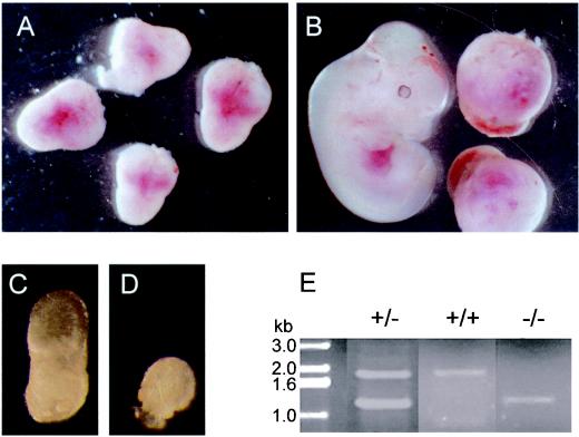
FIG. 2.
(A and B) Embryos dissected at (A) 7.5 dpc and (B) 9.5 dpc from SDHD+/− females mated with SDHD+/− males. Note differences between embryos in panel B, which are not observable in panel A. (C and D) Embryos dissected from maternal decidua at 7.5 dpc. (E) PCR analysis of embryos dissected at 7.5 dpc. Stalled embryos show only the band corresponding to the mutant SDHD allele (−/−; 1.25 kb), whereas normal embryos show the pattern expected for either wild-type (+/+; 1.8 kb) or heterozygous (+/−; both bands) individuals.

